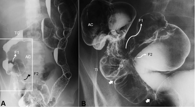
Figure 4. Double-contrast barium enema study. Entire picture of the colon (A) and enlarged picture of the ileocolic portion (B). A – Shortening of the right colon is seen. Diffuse coarse mucosa from the sigmoid colon to the distal part of the descending colon is observed. B - Two stenotic sites in the terminal ileum are indicated by white arrows. There are two entero-enteric fistulas indicated by F. TC, transverse colon; AC, ascending colon; TI, terminal ileum; F, fistula.

