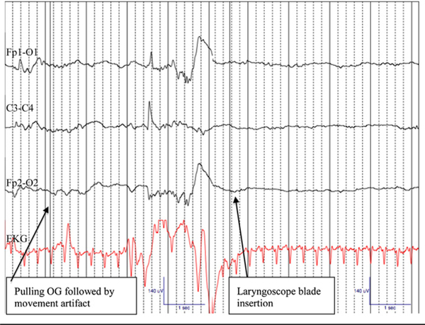
Figure 2.
Amplitude suppression and background pattern changes on EEG Channels: Fp1-O1, C3-C4, Fp2-O2, EKG following laryngoscope blade insertion for endotracheal intubation in a 31 week female (1855 g) premedicated with atropine, narcotic, and paralytic prior to endotracheal intubation for respiratory distress syndrome. Artifact noted on EEG following removal of orogastric tube and just before laryngoscope blade insertion. Gain: 7 mm, Low Frequency Filter of 1 Hz, High Frequency Filter of 20 Hz. Recording speed 30 mm/ sec. OG, orogastric tube.

