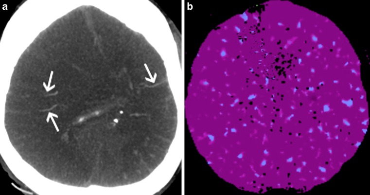
Fig. 4.
Results of CTA (a) and CTP (b) in the patient diagnosed with brain death. CTA shows filling of cortical branches of the right and left MCA (arrows) and was classified as negative, i. e. inconsistent with the diagnosis of BD. CTP reveals perfusion values below the thresholds for non-viable tissue and, contrary to CTA was interpreted as positive, i. e. consistent with the diagnosis of BD

