Fig. 6.
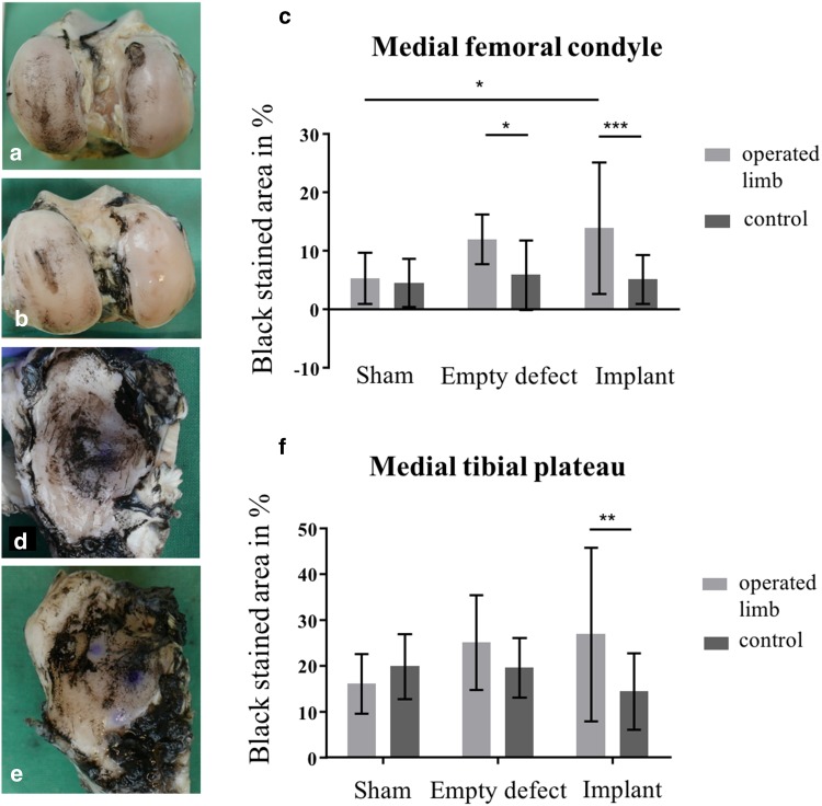
India ink staining of the articular surfaces of femur and tibia. a Femoral condyles of an animal of the meniscectomy group, displaying considerable staining at the medial femoral condyle. b Femoral condyles of an animal of the implant group. c Percentage (%) of black stained area at the medial femoral condyle. Mean ± standard deviation; *p < 0.05; ***p < 0.001. d Medial tibial plateau of an animal of the meniscectomy group, displaying extended fibrillation of the cartilage surface. e Medial tibial plateau of an animal of the implant group. f Percentage (%) of black stained area at the medial tibial plateau. Mean ± standard deviation; **p < 0.01

