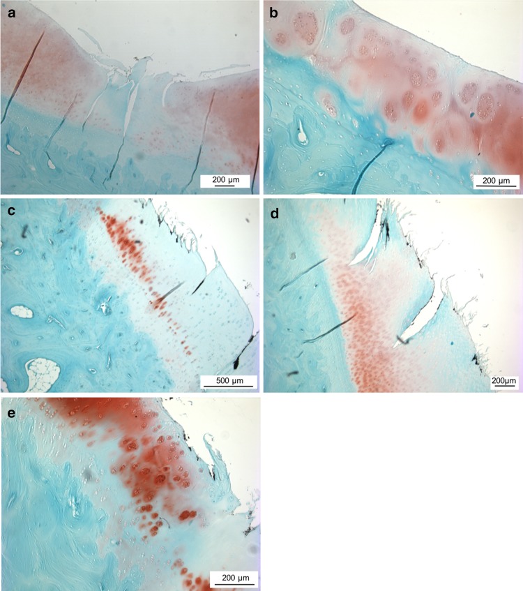
Fig. 7.
Histological images of the articular cartilage of the medial tibial plateau. a Histological section of the medial tibial plateau of an animal of the meniscectomy group, displaying a considerable reduction in safranin-o staining intensity, fissures down to the calcified cartilage as well as diffuse hypocellularity. 50 ×-magnification. b Magnification of a. Clusters of chondrocytes. 100 ×-magnification. c Tibial cartilage after meniscal replacement, showing considerable loss of staining intensity as well as reduction in cellularity. 50 ×-magnification. d Fissuring of cartilage surface and cracks into the radial zone of articular cartilage after meniscal replacement. 50 ×-magnification. e Magnification of c. Chondrocytes arranged in clusters. 100 ×-magnification

