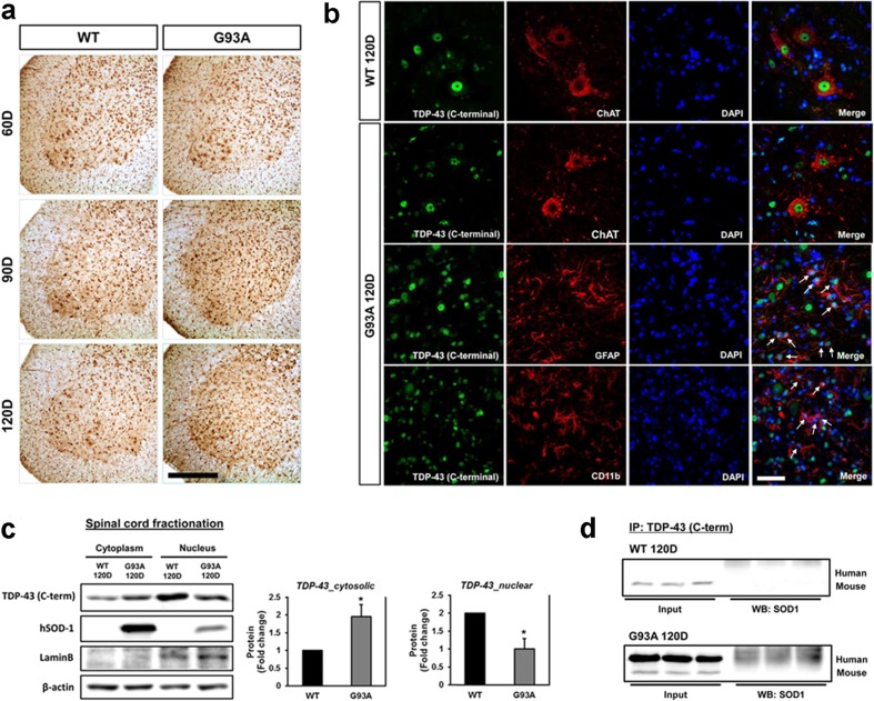
Fig. 2.
TDP-43 (C-terminal) immunoreactivity increased in the spinal cords of hSOD1G93A mice. a Representative images of TDP-43 immunostaining in the ventral horn of lumbar spinal cord sections from WT and hSOD1G93A mice at 60, 90, and 120 days of age. A significant increase in the immunoreactivity of TDP-43 in non-neuronal cell was observed at 90 and 120 days of age in comparison to control mice. b TDP-43-positive astrocytes (GFAP+) (arrows) or TDP-43-positive microglial cells (CD11b+) (arrows). Scale bars: 10 μm. c Significantly higher levels of TDP-43 in the cytosolic fraction was observed in 120-day-old hSOD1G93A mice than in the age- and sex-matched WT mice. d We confirmed that SOD1-TDP-43 interactions were significantly increased in 120-days-old SOD1G93A mice of lumbar spinal cord. Values are mean ± SEM, n = 6; *P < 0.05

