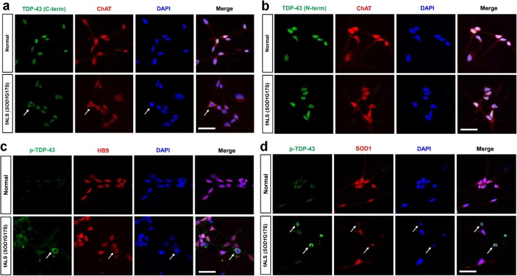
Fig. 7.
Localization of TDP-43 in iPSCs-derived motor neurons from SOD1G17S fALS patient. Representative micrographs at confocal microscopy using three TDP-43 antibodies. a, b The normal shows intense TDP-43 (C-terminal and N-terminal) staining in all nuclei. a Diffuse cytoplasmic mislocalization of TDP-43 (C-terminal) colocalized with ChAT positive motor neurons (arrows). b TDP-43 (N-terminal) only localized with nucleus of ChAT positive motor neurons. c, d p-TDP-43-positive inclusions colocalized with SOD-1-immunoreactive inclusions in the motor neuronal cytoplasm (arrows)

