Fig. 6.
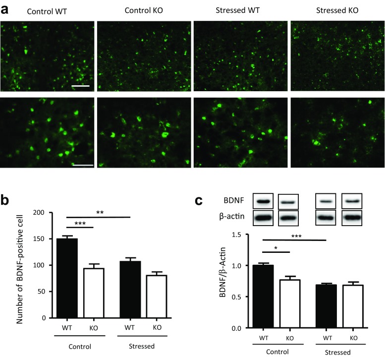
Effects of stress on the expression of brain-derived neurotrophic factor (BDNF) in the dorsal raphe nucleus (DRN) of modulator of apoptosis 1 (MOAP-1)−/− (KO) and wild-type control (WT) mice (age 3–6 months). a Representative photomicrographs of BDNF immunofluorescent staining in the DRN of the WT and MOAP-1−/− mice with or without stress treatment. Scale bar = 100 μm (top panel) or 50 μm (bottom panel). b Number of BDNF immunopositive cells in the DRN in WT and MOAP-1−/− mice with or without stress treatment, n = 4–5. Data are presented as mean ± SEM. ANOVA: F = 17.38, P < 0.001. **P < 0.01 and ***P < 0.001 against control WT by Bonferroni correction. c Expression of BDNF in the midbrain region by Western blot analysis, n = 3–4. Data are presented as mean ± SEM. ANOVA: F = 10.52, P < 0.05. *P < 0.05, ***P < 0.001 by Bonferroni correction. Representative blot bands of the corresponding groups are shown in the top panel

