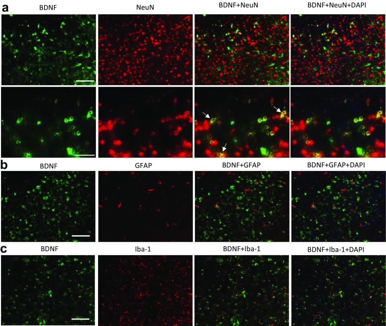
Fig. 7.
Cellular localization of brain-derived neurotrophic factor (BDNF) in the dorsal raphe nucleus (DRN) of wildtype mice. a Double staining of BDNF with NeuN. Scale bar = 100 μm (top) and 50 μm (bottom). White arrows indicate colocalization. b Double staining of BDNF with glial fibrillary acidic protein (GFAP). Scale as in (a), top panel. c Double staining of BDNF with induction of brown adipocytes 1 (Iba-1). Scale as in (a), top panel

