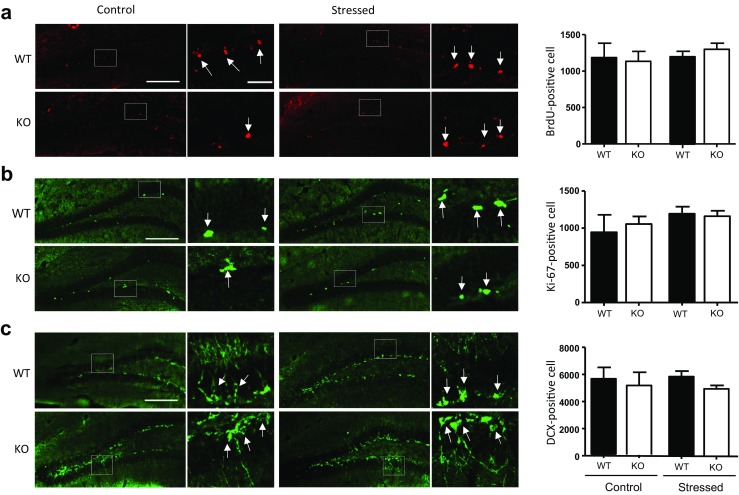
Fig. 9.

Neurogenesis in the hippocampus of modulator of apoptosis 1 (MOAP-1)−/− (KO) and wild-type control (WT) mice following 3d-RFSS (age 3–6 months). Immunohistochemical staining of a BrdU, b Ki67, and c DCX in the dentate gyrus. The right panels are representative photomicrographs. Arrows indicate positively stained cells. The white box indicates the area where the high magnification photomicrograph was taken. Scale bar = 200 or 40 μm. The left panels present the number of positively stained cells. Data are presented as mean ± SEM, n = 4. Statistical analysis performed by one-way ANOVA: a F = 0.2749, b F = 0.6245, c F = 0.3487, no statistical significance
