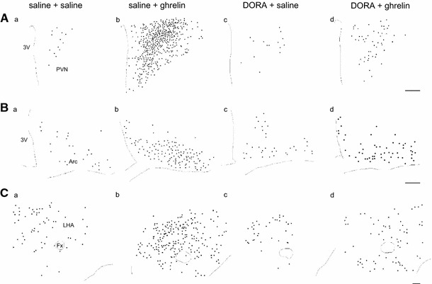
Fig. 2.

Distribution of Fos-immunoreactive (IR) neurons in the paraventricular nucleus (PVN; A a–d), the arcuate nucleus (Arc; B a–d), and the lateral hypothalamic area (LHA; C a–d) at 90 min after icv administration of ghrelin (2 nmol) or saline, pretreated with orexin receptor antagonist (DORA, ACT462206) or saline in the selected sections. Circle dots indicate the localization of the neurons in which Fos-IR was observed. 3V third ventricle, Fx fornix. Scale bars indicate 100 μm
