Fig. 3.

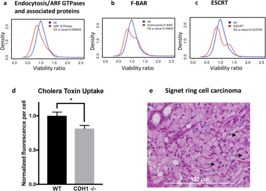
Density distributions of CDH1−/−/MCF10A viability ratios for a endocytosis/ARF GTPases and associated proteins, b F-BAR curvature proteins and c ESCRT curvature proteins. d Differential uptake of fluorescently labelled cholera toxin subunit B in MCF10A and CDH1−/− cells. Cells were grown in medium deprived of cholera toxin (a standard component of MCF10A growth media) for 48 h before addition of Alexa Fluor 488-labelled cholera toxin B. After 30 min, cells were washed and fixed before cell counting and measurement of fluorescence intensity. Fluorescence was normalised to both the total cell count and the MCF10A result. e Haematoxylin and eosin stain of gastric stage T1a carcinoma from a germline CDH1 mutation carrier showing mucin-filled signet ring cells. Three examples are indicated with black arrows
