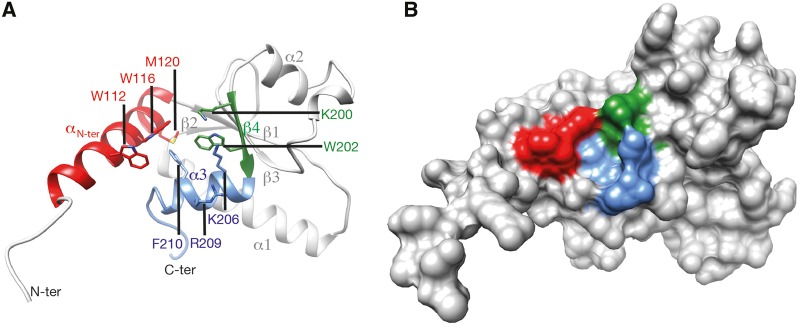
Fig. 5.
—The H-BRCT domain 3D structure. Experimental 3D structure of the H-BRCT domain of S. cerevisiae Dbf4 (PDB 3QBZ; Matthews et al. 2012), on which are highlighted the highly conserved positions depicted with stars in figure 5. (A) Ribbon representation. (B) Solvent accessible surface.

