Figure 1.
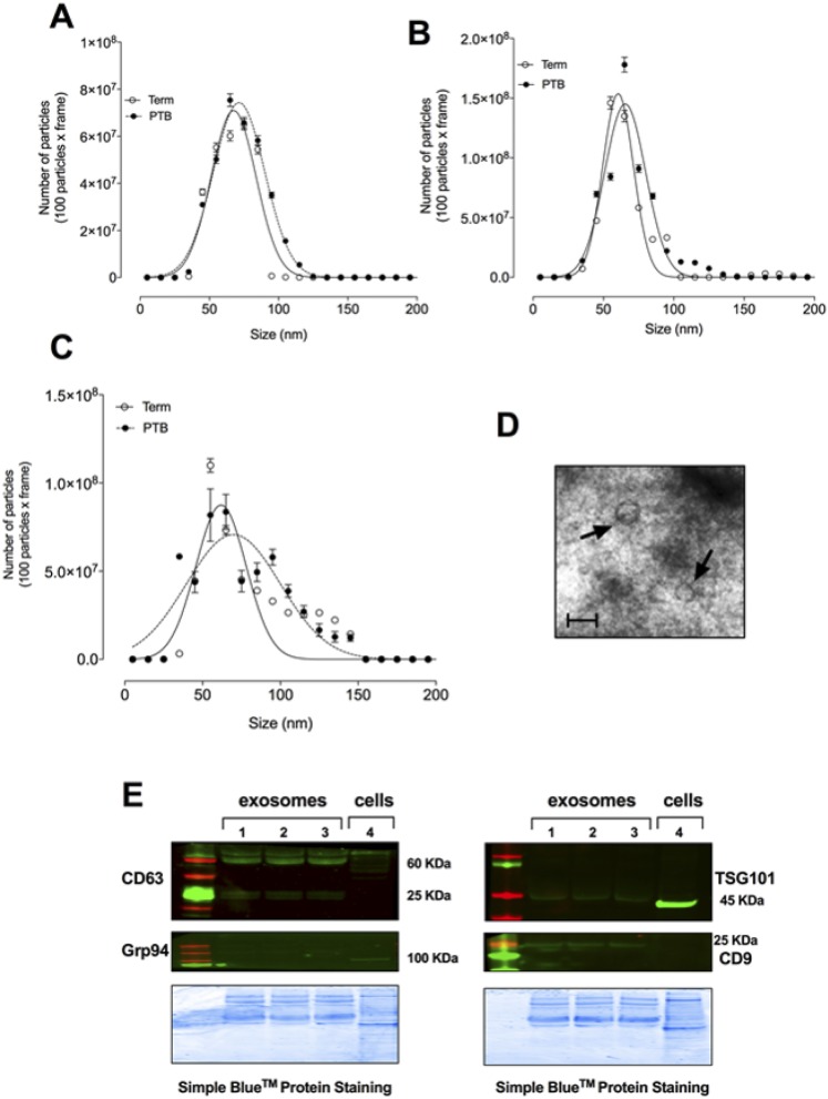
Isolation of exosomes. Exosomes were isolated from maternal plasma obtained from normal and PTB pregnancies across gestation (see “Subjects and Methods”). (A–C) Graphical representation of the vesicle size distribution using a NanoSight NS500 instrument at different time points during pregnancy [(A) first trimester, (B) second trimester, and (C) third trimester]. (D) Representative image of electron micrograph of exosomes. Scale bar, 100 nm. (E) Representative Western blot for enriched exosome markers CD63, CD9, and TSG101, as well as negative control Grp94 for exosomes isolated from normal and PTB at different time points during pregnancy. In (A)–(C) and (E), none of the experiments performed was significantly different for normal vs PTB.

