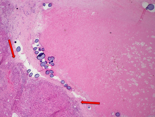
Fig. 6.

Microvalve infusion explant specimen. There is extensive fibrotic tissue identified compatible with necrotic tumor with a small region of viable tumor tissue (red arrows) still present in this specimen demonstrating 90% tumor necrosis. Several groups of drug-eluting microspheres are also noted within the tumor bed
