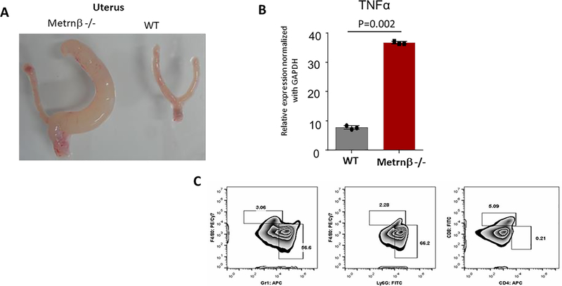
Figure 9. Recurring lesions observed in uterus of Metrnβ−/− mice show inflammation.
(A) Pathology observed in one of the horns of a uterus from an Metrnβ−/− mouse compared with WT littermate uterus. (B) TNFα upregulation in Metrnβ−/− mouse uterus was measured by qPCR. (C) Flow cytometry analysis revealed leukocytic infiltrate, including neutrophils (Gr1; Ly6G) and macrophages (F4/80). Bars represent mean +/− SEM. ****p<0.005; Results are representative of at least 3 experiments.

