
Figure 6:
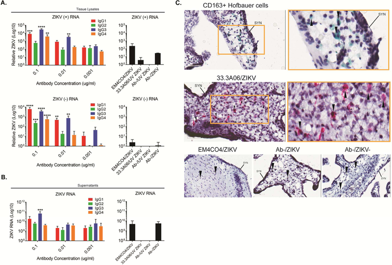
IgG1 and IgG3 subclasses preferentially enhance ZIKV infection of human placental explants. A) Human placental explants were infected with ZIKV (5×10⁵ PFU/ml) in the presence of 33.3A06 mAb IgG subclasses (0.1, 0.01, 0.001 µg/mL). Viral replication within tissues was assessed by strand-specific qRT-PCR at 24 hpi (biological triplicates ±SD). Control conditions used ZIKV alone and UV-ZIKV at 5×10⁵ PFU/ml and mAbs at 0.1 ug/ml. Representative experiment from n=3 donors. B) Supernatants from human placental explants infected as in (A) were collected at 24 hpi and ZIKV RNA measured by qRT-PCR (biologic triplicates). N=2 donors. Data were analyzed by 1-way ANOVA and Dunnett’s multiple comparison test comparing log-transformed ZIKV RNA with 33.3A06 IgG subclass ZIKV RNA levels to the log-transformed non-specific antibody control, *p<0.05, **p<0.01, ***p<0.001, ****p<0.0001. C) Explants were infected with ZIKV (5×10⁵ PFU/ml) in the presence of 33.3A06 (0.4 ug/ml) or EM4CO4 (0.4 ug/ml). HCs (top) and ZIKV E protein (middle and bottom) were visualized by chromogenic staining with anti-CD163 and anti-4G2 antibodies, respectively. Magnification is at 40x (left) and 80x (right, yellow box). Controls are shown across the bottom (40x magnification). SYN, syncytiotrophoblast layer. Arrows indicate HCs. N=2 donors.