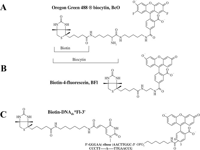
Fig 1. Dye-labeled B7 probes.
(A) Biotin-4-fluorescein (BFl) contains a shorter spacer of 10 non-hydrogen atoms between the bicyclic ring and the dye structure. (B) Oregon green 488 Biocytin (BcO) has a spacer of 20 non-hydrogen atoms between the bicyclic ring and the fluorescent dye. Biocytin (Bc) is an amide formed with B7 and L-lysine. (C) biotinylated DNA labeled at the 3’ end with fluorescein (B7-DNAds*Fl-3’), where B7 was attached to a 14-mer DNA duplex labeled with fluorescein (Fl) at the 3’ end with 16 non-hydrogen atoms between the bicyclic ring and the thymine cyclic base. Unlabeled B7 was used to find the reaction rate of the final binding site in AV and compare it with the reaction rates of the initial binding site to assess possible cooperativity.

