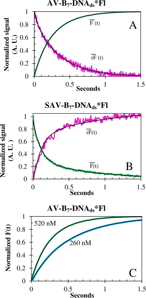
Fig 7. Association traces of biotin-DNAds*Fl binding to SAV and AV.
The and signals of the association reactions of B7-DNAds*Fl (20nM) to (A) AV (520 nM) and (B) SAV (200 nM), at 15°C. (C) Concentration dependence of B7-DNAds*Fl (20 nM) binding to AV at 15°C. All curves (black line) were strongly biphasic. Notice the inversion of SF signals. However, the traces were in prefect agreement with QY experiments (Table 4).

