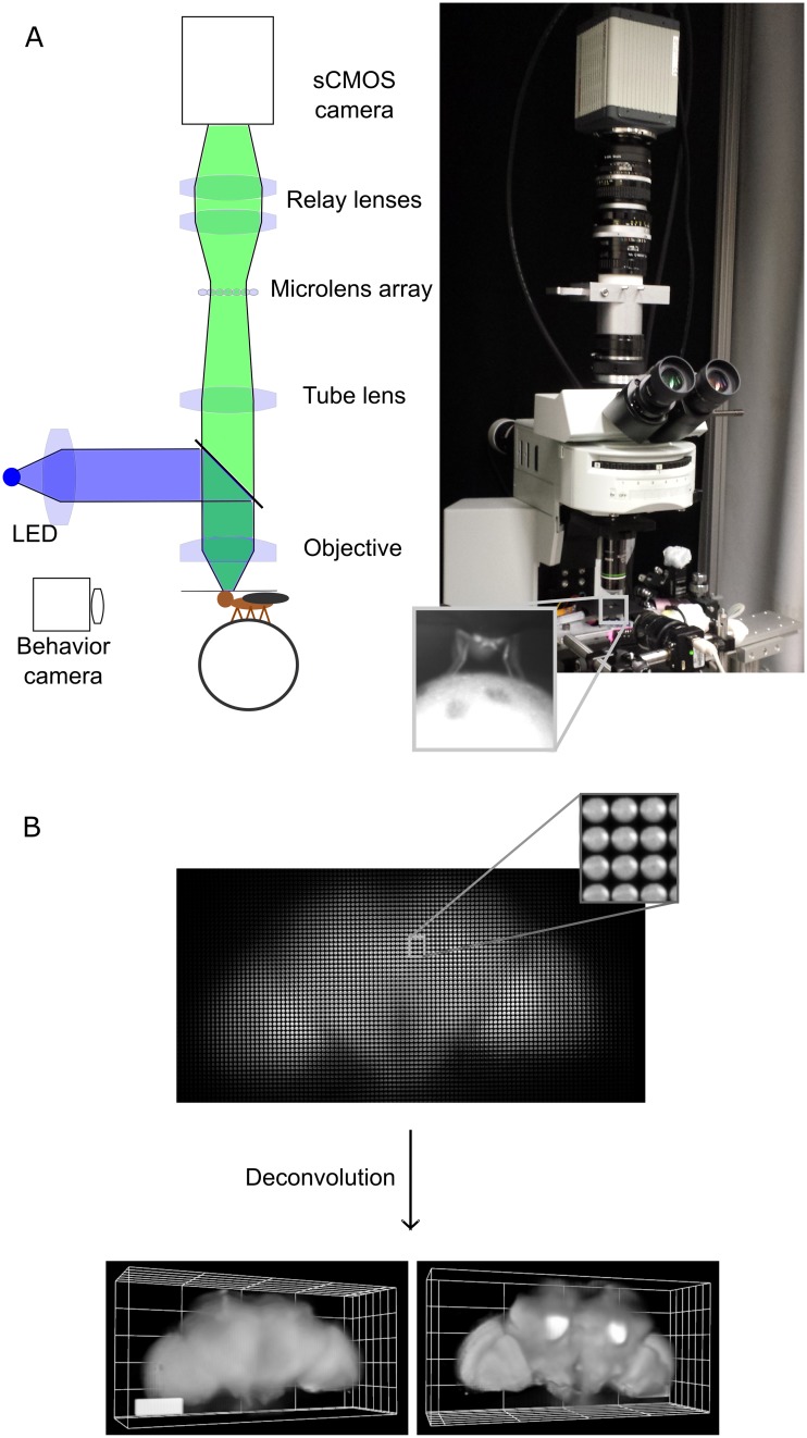
Fig 1. Imaging the brain of adult behaving flies using light field microscopy.
A) Experimental setup. The fly is head fixed, and its tarsi are touching a ball. The light from the brain’s fluorescence goes through the objective, the microscope tube lens, a microlens array, and relay lenses onto the sensor of a high-speed sCMOS camera. Another camera in front of the fly records its behavior. B) Example of a light field deconvolution (fly’s genotype: nsyb-Gal4, UAS-ArcLight). Top: 2D light field image acquired in 5 ms—one camera acquisition period—with a 20x NA 1.0 objective. Bottom: anterior and posterior views (slightly tilted sideways) of the computationally reconstructed volume. 3D bar is 90 x 30 x 30 microns. See also S1–S3 Figs. NA, numerical aperture; nysb-Gal4, nSynaptobrevin-Gal4; sCMOS, scientific complementary metal-oxide-semiconductor.

