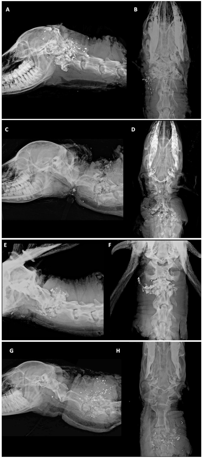
Fig 2. Orthogonal digital radiographs of penetrating ballistic injury in a deer with C1-3 shot placement.
Orthogonal digital radiographs of penetrating ballistic injury in a deer shot with the point of aim targeted at C1-3 (A), (C), (E) lateral; and (B), (D), (F) ventrodorsal. The cranial cervical spine is the point of impact. Regional destruction is noted. Numerous amorphous, distorted (submillimeter to <2 cm) ballistic lead fragments overlie the resultant skull and C1-2 bone fragments. Bone fragments have a “pulverized” appearance resulting from combined ballistic kinetic features related to velocity, mass, and surface area, and the rotational forces (or tumbling) imposed on the projectile when it impacts the target. Notice the comminuted, lucent fracture lines (arrows) coursing rostrally through the calvarium and fracture of the occiput, occipital condyle, and petrous temporal bones. This demonstrates the explosive nature of the impact. There is extensive fragmentation and disruption of the normal architecture of C1 and C2, the vertebral spinal canal alignment and the normal anatomic relationships of the atlanto-occipital junction to C2. Numerous amorphous ballistic lead fragments overlie the many bones of the affected cranial cervical spine demonstrating the transfer of kinetic energy to the tissues beyond the path of the projectile and the related collateral tissue damage. There is complete disruption of the spinal canal with compression and collapse of C1-2 and associated soft tissue swelling (edema and hematoma formation). As the point of impact moves caudally (at C2 and C3), the caudal calvarium is preserved with deformation and collapse of the vertebral arch, lamina, and intervertebral disc space at C2-3. Note the bullet fragments in the spinal canal at C2, missile fragmentation and the circumferential destruction and obliteration of the spinal canal. Although not visible, the spinal cord was also obliterated (confirmed on post-mortem evaluation). In other cases of cranial cervical point of impact, traumatic disarticulation (subluxation/luxation) was observed. The soft tissue destruction can extend ventrally to the laryngeal soft tissues resulting in gas tracking through the deep fascial planes of the neck, and pharyngeal and esophageal perforation. In (G) and (H), the gas throughout the length of the esophagus (asterisk) likely resulted from post-mortem tissue handling.

