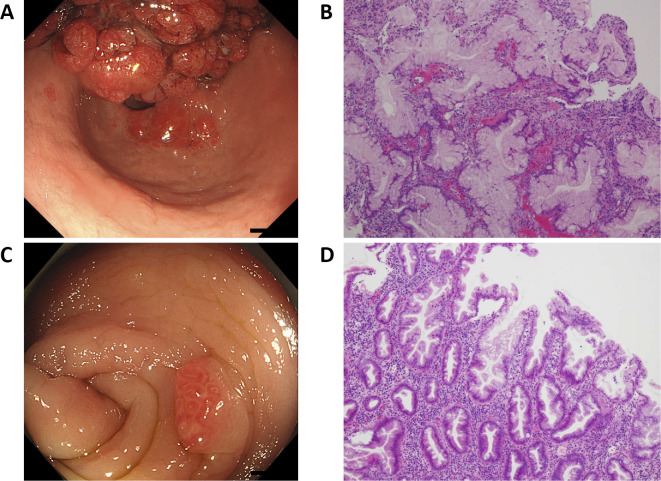
Figure 1.
Gastrointestinal endoscopy and colonoscopy. A: The gastric polyps were massive and bled easily. B: Hematoxylin and Eosin (H&E) staining of the gastric polyps. Foveolar hyperplasia and small round cells were observed (lymphocyte>plasma cell). The gastric foveola showed an increased complexity of structure and density. C: Multiple polyps were located throughout the colon. D: H&E staining of the colon polyps. Hyperplastic polyps of the microvesicular type were observed, along with mild dysplasia of the gland duct.

