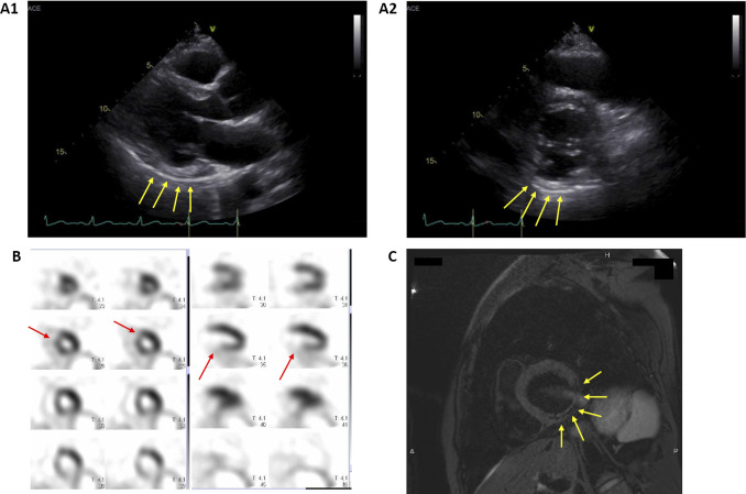
Figure 5.
Imaging findings of the left ventricle (LV). A1 and A2: Echocardiography revealed that the inferior and posterior walls of the LV were hyperechoic and hypokinetic (yellow arrows). The LV ejection fraction was 49%. While mild mitral regurgitation was found, the aortic, pulmonary, and tricuspid valves showed no remarkable findings. B: Myocardial perfusion scintigraphy revealed a perfusion abnormality in the inferior, posterior, and anteroseptal walls of the LV (red arrows). C: Cardiac MRI revealed fatty degeneration of the inferior and posterior walls of the LV, showing a low signal on fat-suppressed T2-weighted imaging (yellow arrows).

