Figure 3.
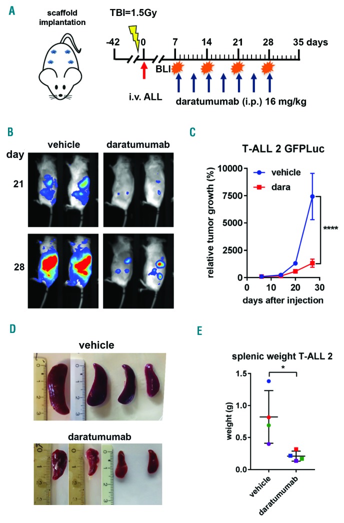
Daratumumab (dara) shows efficacy in a patient-derived xenograft (PDX) mouse model for aggressive T-cell acute lymphoblastic leukemia (TALL) growth. (A) Rag2−/−γc−/− mice carrying scaffolds coated with human mesenchymal stromal cells were inoculated (red arrow) with luciferase-transduced primary acute lymphoblastic leukemia (ALL) cells of T-ALL patient 2 (T-ALL2). Mice were treated with vehicle control or dara at the indicated time points (blue arrows). Mice were monitored weekly by bioluminescent imaging (BLI), and sacrificed at day 33 at which time point splenic weight was determined. (B). BLI at day 21 and 28 of dara treatment. Representative images are shown. (C). Tumor growth was determined by average photon emission intensity measured for a region of interest in time. The results are expressed as relative growth with the intensity at day 6 set to 100. The lines represent the mean signal of 4 mice±Standard Error of mean. ****P<0.001 compared to vehicle, two-way ANOVA, Sidak’s multiple comparison test. (D). Splenic weight was plotted for vehicle and dara-treated mice where the number of dots represents the number of mice per treatment group. *P<0.05 vehicle, Mann-Whitney test. TBI: total body irradiation; i.v.: intravenous injection; i.p.: intraperitoneal; g: gramms.

