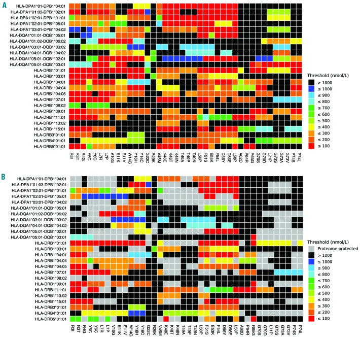
Figure 5.
MHC-binding strengths of F8 peptides predicted to form novel pMHC surfaces with and without proteome scanning. Heatmap showing the predicted occurrence of novel pMHC surfaces and binding strengths for 25 HLA-DR/DP/DQ isoforms (y axis) covering the first 50 missense mutations in the Factor VIII Gene (F8) Variant Database (x axis). Black and gray squares indicate F8 missense mutation/HLA isoform combinations that are not predicted to form a novel pMHC surface. Otherwise the temperature color scale indicates the predicted binding strength of the strongest binding peptide with a novel pMHC surface for each remaining F8 missense mutation/HLA isoform combination. The full heatmap for all missense F8 mutations is given in Online Supplementary Figure S1. (A) MHC-binding strengths of F8 peptides predicted to form novel pMHC surfaces (colored squares), or not (black squares), without proteome scanning. (B) MHC-binding strengths of F8 peptides predicted to form novel pMHC surfaces with proteome scanning. Gray squares indicate F8 missense mutation/HLA isoform combinations that are no longer predicted to form a novel pMHC surface after cross-matches to the proteome are taken into account. MHC: major histocompatibility complex; pMHC: peptide-major histocompatibility complex; HLA: human leukocyte antigen.

