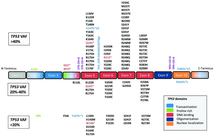
Figure 1.
Distribution of 108 TP53 mutations found in diagnostic specimens of 98/1537 patients with acute myeloid leukemia. Top panel: TP53 mutations with a variant allele frequency (VAF) of >40%; middle panel: mutations with a VAF of 20%-40%; lower panel: mutations with a VAF <20%. Missense mutations are marked in black, nonsense in red, insertions/deletions in blue and essential splice site mutations in purple. Despite different VAF, the vast majority of TP53 mutations are missense mutations located within the DNA binding domain of the gene.

