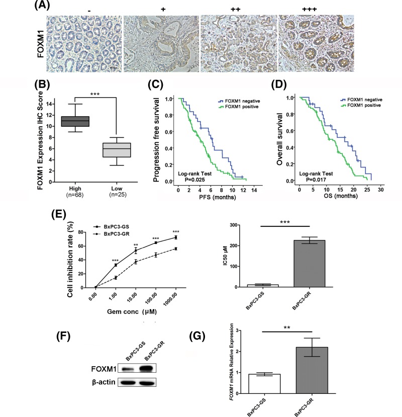
Figure 1. FOXM1 expression is associated with gemcitabine resistance in pancreatic cancer.
(A) Representative images of FOXM1 protein expression in paraffin-embedded tissue from 93 patients with pancreatic cancer (−, +, ++, +++). (B) According to the immunohistochemical score, the patients were divided into a high-FOXM1 expression group (n=68) and a low-FOXM1 expression group (n=25). (C) The PFS curves for the high-FOXM1 expression group and the low-FOXM1 expression group. The PFS rates between the two groups were significantly different (P=0.025). (D) The OS curves for the high-FOXM1 expression group and the low-FOXM1 expression group. The difference is statistically significant (P=0.017). (E) BxPC3-GS and BxPC3-GR cells were treated with different concentrations of gemcitabine for 48 h. Cell viability was determined using CCK-8 assays. (F) Western blotting analysis to determine the relative protein levels of FOXM1 in BxPC3-GS and BxPC3-GR cells. (G) Relative mRNA expression levels of FOXM1 between BxPC3-GS and BxPC3-GR cell lines were compared by qPCR. Overexpression of FOXM1 mRNA was confirmed in BxPC3-GR cells. Scale bar, 100 μm. **P<0.01; ***P<0.001.

