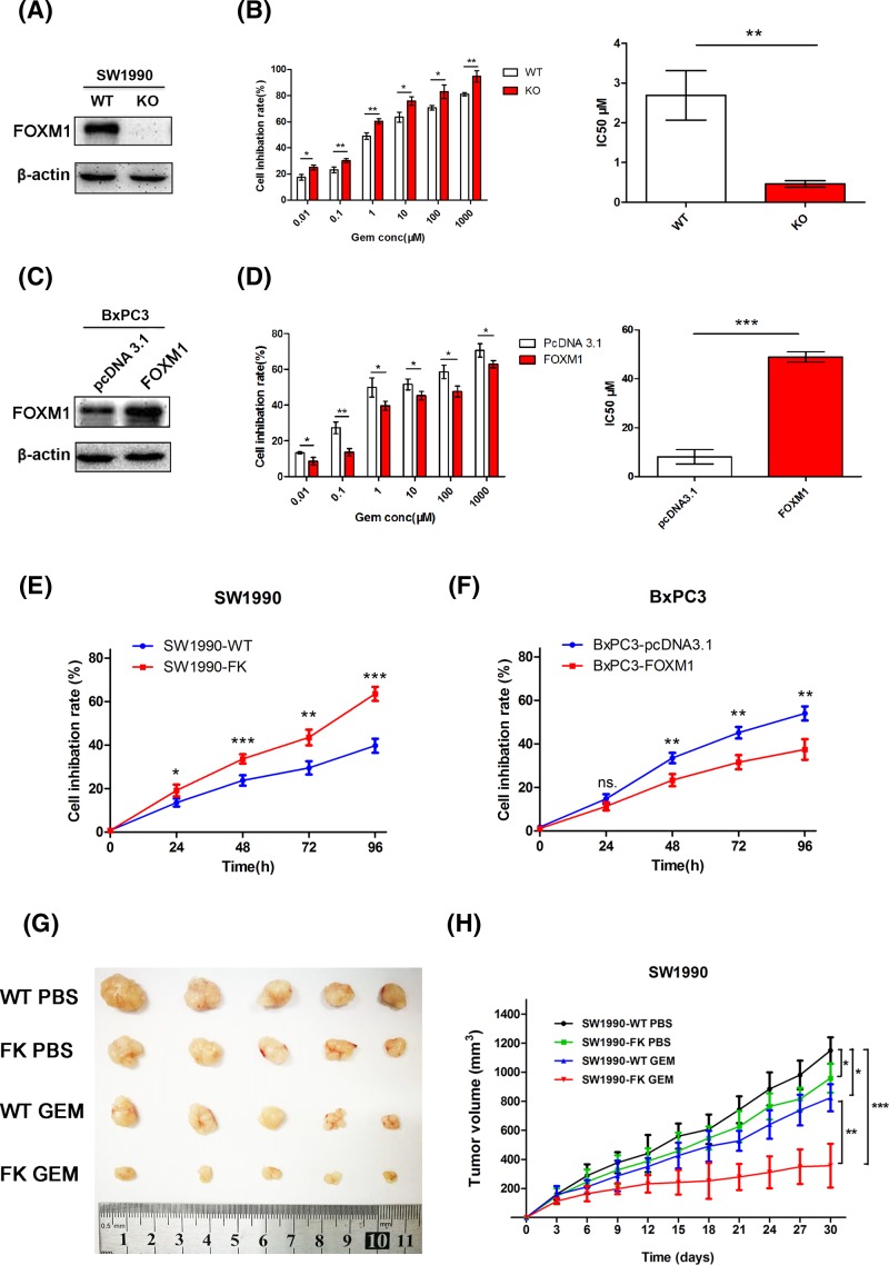
Figure 2. FOXM1 decreased the sensitivity of pancreatic cancer cells to gemcitabine in vitro and in vivo.
(A,C) Western blotting analysis to determine the relative protein levels of FOXM1 in SW1990-WT/FK, BxPC3-pcDNA3.1/FOXM1 cells. (B,D) CCK-8 assays were performed to detect the viability these cells treated with varying concentrations of gemcitabine for 48 h and to calculate the IC50. (E,F) SW1990-WT/FK and BxPC3-pcDNA3.1/FOXM1 cell lines were treated with DMSO or 100 nM gemcitabine for 4 days and cell inhibition rate at each time point was measured with CCK-8. (G,H) SW1990-WT/FK cells were subcutaneously injected into the left flank of nude mice. Administration of chemotherapy began when the tumor diameter reached 3–5 mm. The mice were randomly divided into four groups (n=5) and treated as described in figure. (G) Tumor size was shown after approximately 30 days of treatment. (H) Tumor volumes were measured every 3 days. A tumor growth curve was drawn according the measured tumor volume. *P<0.05; **P<0.01; ***P<0.001.

