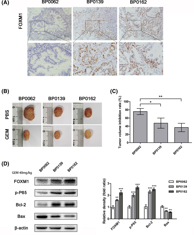
Figure 3. Pancreatic cancer tissues with high expression of FOXM1 are less sensitive to gemcitabine in PDX tumors.
(A) In three pancreatic cancer PDX models, FOXM1 expression levels were analyzed by IHC. Scale bar, 100 μm. (B) These PDX tumor sizes were shown after grouping and treatment for 3 weeks. (C) The inhibition rate of gemcitabine treatment for each PDX model. (D) The expression of FOXM1, p-P65, and apoptotic proteins Bcl2 and Bax in the tumor tissues after treatment was detected by Western blotting. *P<0.05; **P<0.01; ***P<0.001.

