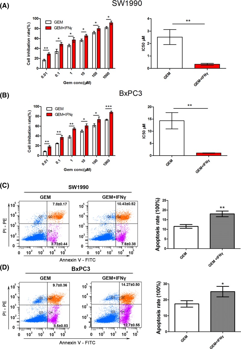
Figure 7. IFNγ can facilitate gemcitabine-induced cell apoptosis.
(A,B) CCK-8 assays were performed to detect SW1990 and BxPC3 cell viability after treatment with varying concentrations of gemcitabine ± 100 ng/ml IFNγ for 48 h and to calculate the IC50. (C,D) After treatment with 100 nM gemcitabine ± 100 ng/ml IFNγ for 24 h, the apoptosis rate of SW1990 and BxPC3 cells was calculated using flow cytometric determination of Annexin V and PI staining. Upper-right + lower-right quadrants indicate apoptotic cells. Apoptosis is quantitated in the bar graphs for SW1990 (top) and BxPC3 (bottom). *P<0.05; **P<0.01; ***P<0.001.

