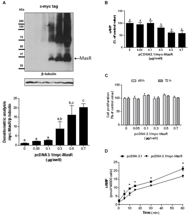
Figure 1.

Constitutive activity of MasR in HEK293T transfected cells. (A) Analysis of MasR expression by Western Blot. Cells were transfected with the indicated amounts of a plasmid encoding the c-myc tagged MasR (pcDNA3.1/c-myc-MasR) and cell extracts were resolved by SDS-PAGE and Western Blot using anti-c-myc antibody. Detection of β-tubulin was used as a protein loading control. Blots were subjected to densitometry analysis using ImageJ software. Data are presented as mean ± SEM respect to control of at least three independent experiments. Images are representative of at least three independent experiments. Different letters denote significant difference at p < 0.05. (B) cAMP accumulation. Cells were transfected with the indicated amounts of pcDNA3.1/c-myc-MasR and cAMP levels were determined after incubation for 30 min with IBMX. 100% corresponds to cAMP levels in cells transfected with mock (pcDNA3.1). Data represent mean ± SEM of four independent experiments. Different letters denote significant difference at p < 0.05. (C) Cell proliferation. Cells transfected with the indicated amounts of pcDNA3.1/c-myc-MasR were cultured for 48 h or 72 h. Cell viability was determined by MTS assay and reported as a percent of proliferation respect to mock transfected cells (n = 3). (D) Time-course production of cAMP. Cells transfected with 0.5 μg/well pcDNA3.1/c-myc-MasR were stimulated during the indicated times with 1 μM forskolin in the presence of IBMX 1 mM and cAMP levels were determined. Data represent mean ± SEM of three independent experiments. Asterisk denote significant difference at p < 0.05 by plasmid type within the same time point.
