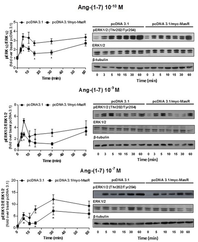
Figure 5.

Kinetics of Ang-(1–7)-induced ERK1/2 phosphorylation. HEK293T cells transfected as described in Figure 4, were incubated with Ang-(1–7) at the different concentrations indicated for the indicated periods. Phosphorylation levels of ERK1/2, total ERK1/2, and β-tubulin were determined by Western Blotting. Phosphorylation of ERK1/2 was normalized to that of total ERK1/2 and expressed as relative to the value obtained at 0 min in cells transfected with the empty vector (pcDNA 3.1). Data represents the mean ± SEM of three independent experiments. ∗P < 0.05 by plasmid type within the same time point.
