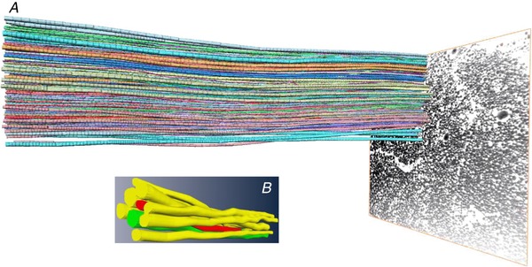
Figure 1. The principle load transmitting structure of tendons.

A, an illustration of tracing of collagen fibrils from a human patellar tendon obtained with FIB‐SEM on a nanoscale level, suggesting that the fibrils are continuous. B, close‐up of individual fibrils that typically range between 30 and 200 nm in diameter. For further detail, see Svensson et al. 2017.
