
Figure 1. Schematic diagram of articular cartilage.
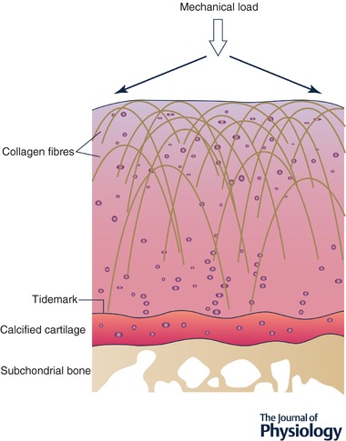
Illustration of tertiary structure of fibrillar collagens in articular cartilage showing parallel arrangement in superficial layer and perpendicular orientation in deeper layers. Arrows show direction of mechanical load and how it is dissipated across the articulating surface.