
FIGURE 4.
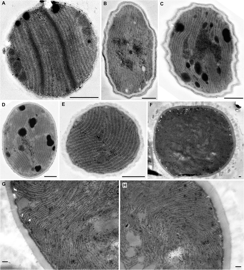
TEM documentation of parallel (A–E) and Cyanothece-like (F–H) thylakoids. A single array of parallel thylakoids filling the entire cell is visible in (A) Cyanobacterium stanieri PCC 7202 and (B–E) Geminocystis papuanica PAP1. (F–H) Thylakoids perpendicular to cell wall protrude into the cell center to form fascicular reticulate structures in Cyanothece aeruginosa SAG 87.79. Scale bars = 500 nm.