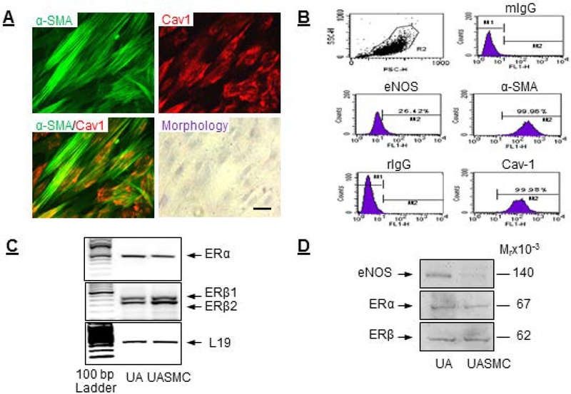
Fig. 1: Establishment and characterization of an ovine uterine artery smooth muscle cells (UASMC) model.
Primary UASMCs from late pregnant ewes were isolated and characterized. (A) Cellular expression and co-localization of α-SMA and caveolin-1 by immunocytochemistry and assessment of cell morphology. (B) Flow cytometry for cellular expression of α-SMA, caveolin-1, and eNOS. ERα and ERβ (C) mRNA and (D) protein expressions in UASMCs and intact UAs. Scale bar is 50 μm.

