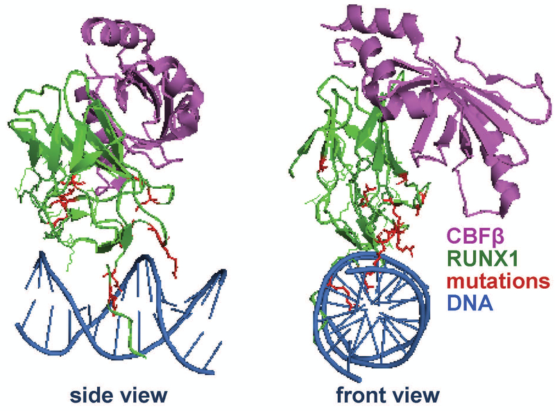
Figure 2. Mutations in the Runt Homology Domain of RUNX1 in breast cancer.
Ribbon representation showing CBF-β in purple, DNA binding Runt Homology Domain in green, and DNA in blue. RUNX1 mutations identified in breast cancer patients are shown in red. For clarity, the structure is shown in two different orientations (front and side), rotated 90 degrees relative to one another. The image was rendered from the Public Data Base (PDB) code 1H9D (Bravo, Li, Speck, & Warren, 2001). The locations of the mutations are in the DNA binding domain suggesting RUNX1 loses its putative function in breast tumors.

