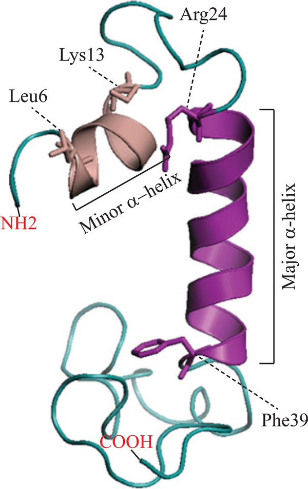
FIGURE 3.
The 3D structure of the full‐length JC virus agnoprotein was recently resolved by NMR (Coric et al., 2017). According to this 3D structure, agnoprotein contains a minor and a major α‐helix located at amino acid position Leu6‐Lys13 and Arg24-Phe39, respectively. The rest of the protein adopts an intrinsically unstructured conformation (Met1‐Gln5, Val14‐Lys23, and Cyt40-Thr71). 3D: three dimensional; NMR: nuclear magnetic resonance [Color figure can be viewed at wileyonlinelibrary.com]

