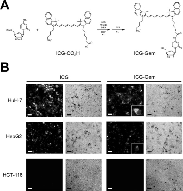
Figure 1.
Chemical structure and fluorescent distribution of an indocyanine green (ICG)-conjugated anti-cancer drug. (A) ICG is conjugated with gemcitabine (Gem) through coupling reactions, followed by deprotection. (B) Fluorescent and bright image (x100) of hepatocellular carcinoma (HCC) cells and colon cancer cell (HCT116), 24 h after exposure to ICG or ICG-Gem. Scale bar = 50 μm. The right lower panel shows the magnified fluorescent image of HCC cells 24 h after exposure to ICG-Gem.

