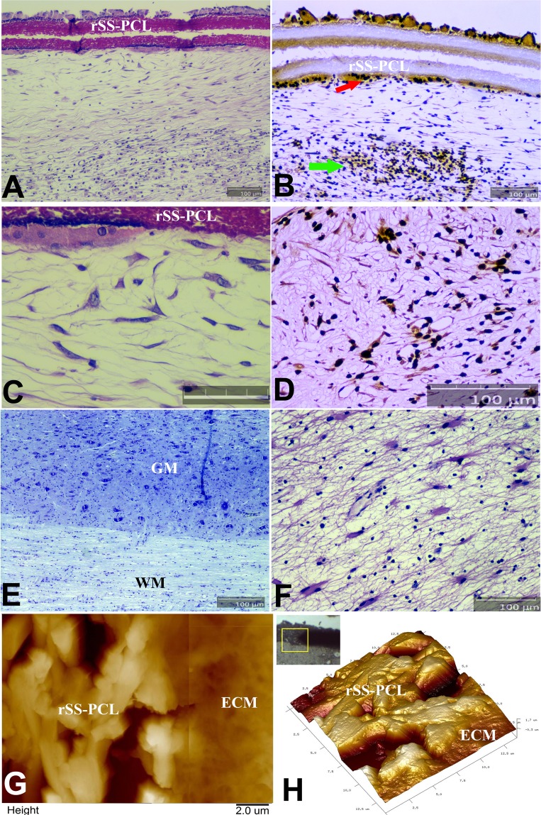
Figure 8.
Histological and immunohistochemical (IHC) analysis of the SPRPix matrix with human drNPC implant with AFM imaging in the projection of the posterior column of the spinal cord of the rhesus macaque. (A) Sagittal section of the spinal cord stained with H&E. (B) Same spinal cord fragment, stained with antibodies to macro-H2A.1 visualized by immunoperoxidase staining. Transplanted cells highlighted by arrows. (C) Enlarged H&E image of (A) demonstrating the cells adjacent to the SPRPix matrix; Bar size = 50 µm. (D) Same image as in (C), stained with antibodies to MAP2. (E,F) Sagittal sections of the spinal cord directly below the SPRPix matrix, Nissl (G) and H&E (H), stains. (G,H) 2D AFM image (overlay of two images from adjacent regions) and a 3D AFM image of the border between the SPRPix and the ECM of the surrounding tissue, showing full integration of the scaffold with the ECM. In panels (A,B,D–F) bar size = 100 µm. Notations: rSS-PCL-spidroin-polycaprolactone scaffold; GM – grey matter; WM – white matter; ECM – extracellular matrix.

