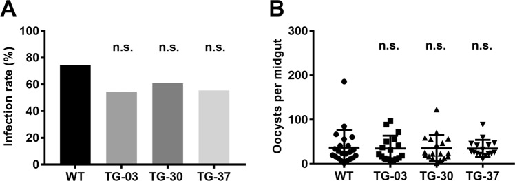
Figure 6.
Sporogony development in TG mosquitoes. (A,B) WT and TG mosquitoes 5–7 days after eclosion were allowed to feed on the same P. berghei-infected mouse. On day 10–12, the midguts (n = 30) were dissected. Infection rate (A) and the number of oocysts (B) formed was counted. Results were analyzed by the Kruskal-Wallis test (A) or Dunnett’s multiple comparisons test (B). Horizontal bars represent the median value. n.s., not significant.

