Figure 1.
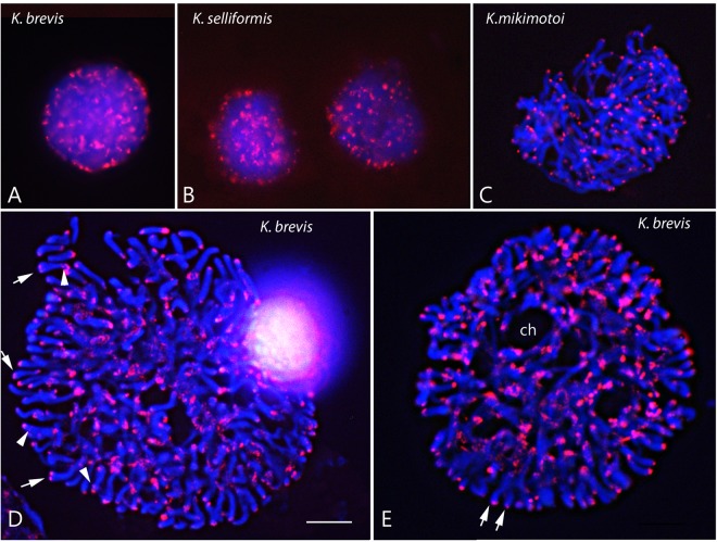
Physical mapping of the telomeric repeats in the nuclei of three Karenia species at different stages of the asexual cycle. K. brevis (A,D,E), K. selliformis (B) and K. mikimotoi (C). Each panel shows merged images, to facilitate visualization of the DAPI-stained DNA (blue) and in situ hybridization of the Dy547-labeled oligonucleotide (CCCTAAA)3 used to localize the telomeric repeats (red) during interphase (A,B) and mitotic stages (C–E). In D, the arrows indicate double visualized telomeric signals, and the arrowheads interstitial signals. The pairs of arrows in E indicate separate sister chromatids. ch = cytoplasmic channels. Scale bar = 10 μm for all panels.

