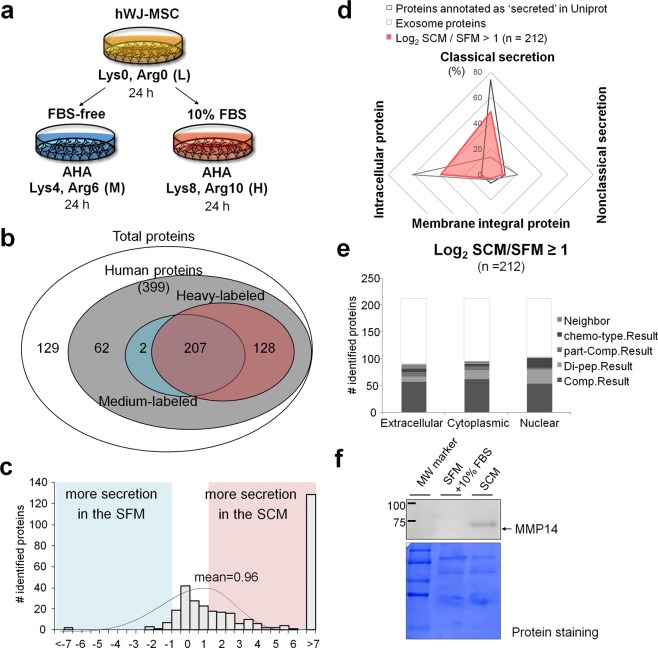
Figure 4.
Analysis of hWJ-MSC secretome from serum-containing medium (SCM) and serum-free medium (SFM). (a) Schematic workflow. (b) The number of identified proteins in the secretome. (c) The distribution of differentially secreted proteins between SFM and SCM. The H/M ratios are log2-transformed after normalization by the difference in growth rate. (d,e) Secretion pathways and subcellular localization of the differentially secreted proteins. (f) MMP14 was measured by western blot analysis. Note that 10% FBS was added to SFM just before SDS-PAGE in order to view any background effect stemming from FBS itself. Equal loading was confirmed by Commassie staining of the membrane. Full-length western blot images are shown in Supplementary Fig. S5b.

