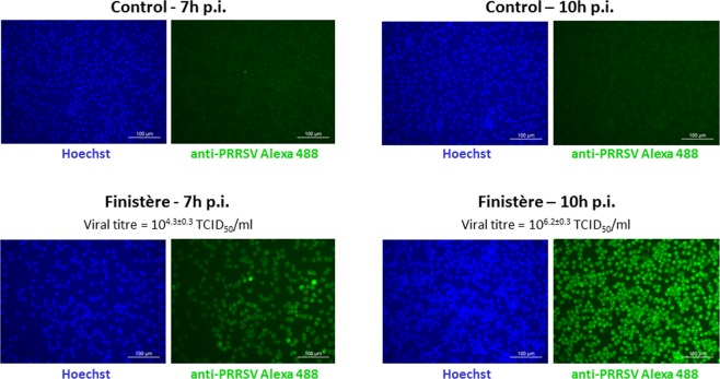
Figure 1.
PRRSV immunofluorescence staining of PAMs infected with the Finistère strain at 7 h and 10 h post-infection (at MOI = 2) and controls. PRRSV indirect staining was performed with anti-PRRSV N protein antibody and anti-IgG Alexa 488-conjugated antibody (green). The nuclei were stained with Hoechst (blue). Magnification: 200X. Images are representative of two biological replicates with three technical replicates for each experimental condition. Viral titers are means ± standard deviations of two biological replicates with two technical replicates for each experimental condition.

