Figure 1.
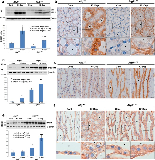
Light micrographs of the inner medulla of control (Cont) and K+-depleted (K+-Dep) Atg7f/f and Atg7Δpc mouse kidneys illustrating immunoblotting (a,c,e) and immunolabeling (b,d,f) of LC3 (a,b), SQSTM1 (c,d) and RAB9 (e,f). (a) The degree of increase in the LC3 II/I ratio is significantly decreased in K+-depleted Atg7Δpc compare to K+-depleted Atg7f/f mice. (b) In the images, even in the control groups, immunoreactivity for LC3 in the inner medullary collecting duct (IMCD, stars) cells are decreased in Atg7Δpc mice compare to Atg7f/f mice. After K+ depletion, immunoreactivity for LC3 in dramatically increased in the IMCD cells of Atg7f/f mice. However, immunoreactivity for LC3 is restrictively decreased in the IMCD cells of K+-depleted Atg7Δpc mice. Note that immunoreactivity for LC3 in other structures including interstitial cells and thin limb cells of Henle’s loop of renal papilla in K+-depleted Atg7Δpc mice is stronger in K+-depleted Atg7f/f mice, but relatively weaker than that of IMCD cells in K+-depleted Atg7f/f mice. (c) The protein level of SQSTM1 is significantly increased in Atg7Δpc compare to Atg7f/f mice. (d) In the images, strong immunoreactivity for SQSTM1 is observed restrictively in the IMCD cells of Atg7Δpc mice, which change is pronounced after K+ depletion. (e) Note the prominent increase of RAB9 in K+-Dep Atg7Δpc mice. (f) Immunoreactivity for RAB9 is not observed in the autophagic vacuoles (arrows) of K+-Dep Atg7f/f mice. However, it is expressed in small autophagic vacuoles of K+-Dep Atg7Δpc mice. Boxes in (b,f) are higher magnification of the areas indicated by the rectangles in upper panels. Values represent the means ± SD.

