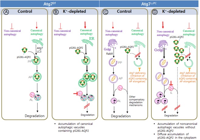
Figure 8.
Schematic diagram summarizing the regulation of canonical autophagy and non-canonical autophagy in pS261-AQP2 degradation, and the cellular accumulation of different autophagic structures in inner medullary collecting duct (IMCD) cells of control (a,c) and K+-depleted (b,d) Atg7f/f (a,b), and Atg7Δpc (c,d) mice. Depicted are the relative amounts of phagophores (PP), autophagosomes (AP), and autolysosomes (AL). (a) Under normal condition of Atg7f/f mice, pS261-AQP2 degradation is regulated by canonical autophagy. There is no accumulation of autophagic vacuoles, because of normal autophagic flux. (b) In K+-Dep Atg7f/f mice, canonical autophagosomes containing pS261-AQP2 are accumulated in IMCD cells as a result of the blockade of autophagic flux by the inactivation of lysosome. Non canonical autophagy is not activated in both control (a) and K+-Dep (b) Atg7f/f mice. (c) When canonical autophagy is suppressed by Atg7 deletion, which is accompanied by enhanced activity of other lysosomal degradation mechanisms. These compensating mechanisms for canonical autophagy deficiency, resulting in no significant accumulation of AQP2 in the IMCD cells of control Atg7Δpc mice. (d) In K+-Dep Atg7Δpc mice, pS261-AQP2 is accumulated throughout the cytoplasm as a result of impairment of compensatory other lysosomal degradation mechanisms by inactivated lysosome. Non-canonical autophagic vacuoles without AQP2 are also accumulated as a result of impairment of non-canonical autophagy flux by inactivated lysosome.

