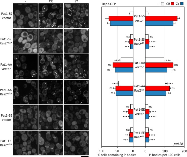
Figure 5.
The presence of elevated PKA signaling activity inhibits P-body induction in response to cell wall stress. pat1Δ cells expressing the Dcp2-GFP protein and transformed with a plasmid that included the constitutively active Ras2val19 or the empty vector (vector) in combination with plasmids including the wild-type Pat1 (Pat1-SS), the non-phosphorylatable variant Pat1-AA or the phosphomimetic Pat1-EE variant were transferred to a medium containing 30 µg/ml CR or 0.8 U/ml ZY for one hour to induce P-body formation. Then, Dcp2-GFP foci were examined by fluorescence microscopy. Representative images are shown for both the control and treatment conditions. The quantitation of the microscopy data, from three independent experiments (n > 100 cells), is shown in the graphs. Statistical significance was determined using a two-tailed, unpaired, Student’s t test by comparing with the no treatment conditions, CR or ZY data from the pat1Δ strain expressing the Pat1-SS version (*P ≤ 0.05, **P ≤ 0.01, ***P ≤ 0.001; ns, not significant). Scale bar, 5 μm.

