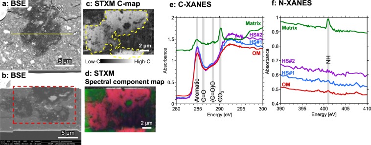
Figure 2.
Scanning transmission X-ray microscopy (STXM) analyses of a focused ion beam (FIB) section containing the organic matter (OM) aggregate in the Zag clast. (a) Backscattered electron (BSE) image of a polished thin section of the organic aggregate (dark) in the carbonaceous clast in the Zag meteorite. FIB section was subsampled from the yellow region. (b) BSE image of the FIB section. STXM maps (c,d) were obtained from the location indicated by the red dotted line. (c) Carbon-map (at 292 eV) indicates the section is dominated by carbon. The OM aggregate is indicated by the yellow dotted line. (d) Spectral component map derived from C-XANES of OM (red) and matrix (green). The red and green regions correspond to the OM and matrix C-XANES spectra shown in (e). (e) The C-XANES of OM aggregate revealed that it is dominated by sp2 carbon (284.8 eV) while in the surrounding matrix carbon is mainly found as carbonates (290.3 eV) with some OM at 286.3 eV that is assigned to ketone (C=O) and 288.5 eV that is assigned to carboxyl/ester [(C=O)O]. (f) The OM aggregate does not show detectable N-XANES features while matrix shows a peak at 401.0 eV which is assigned to amines. The C- and N-XANES obtained from isotope hot spots (HS, see Fig. 3) are also shown.

