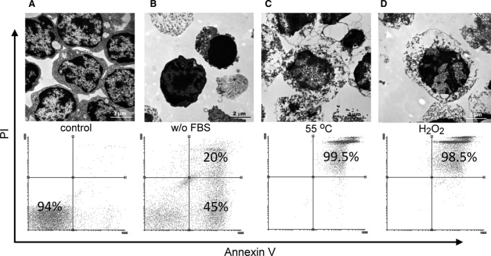
Figure 1.

Morphology and viability of target cells. Transmission electron microscopic images and propidium iodide/annexin V staining of target cells. (A) Thymocytes immediately after isolation are considered as viable cells. (B) Apoptosis induced by serum deprivation for 24 h is characterized with condensed nuclei. According to the propidium iodide staining, both early and late apoptotic cells are included. (C,D) Incubation at 55 °C for 20 min (C) or with 1 mm H2O2 for 24 h (D) induced necrosis with early swelling and propidium iodide positivity. Scale bar: 2 μm.
