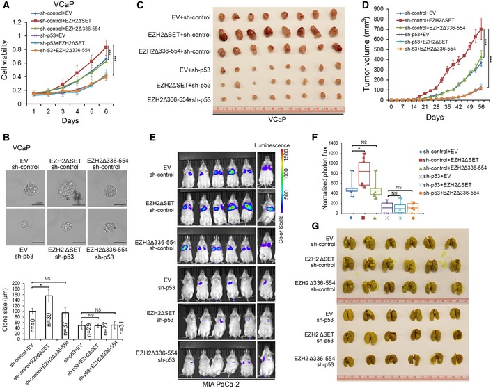
Figure 6. EZH2 enhances p53 GOF mutant‐mediated cancer growth and metastasis independently of its methyltransferase activity.

-
A, BVCaP cells stably expressing control (sh‐control) or p53‐specific shRNA were infected with lentivirus for empty vector (EV) or deletion mutants of EZH2. Cell growth in 2D (A) and 3D (B) conditions were determined by MTS assay and measurement of clone size, respectively. Statistical significance was determined by two‐tailed Student's t‐test. *P < 0.05, ***P < 0.001.
-
C, DVCaP cells (1 × 10⁷) infected with lentivirus as in (A) were injected subcutaneously into NSG mice (n = 8/group). Tumors were measured by caliper twice a week. Tumors at the end point of measurement were isolated and photographed (C), and data are shown as means ± SD (D). Statistical significance was determined by two‐tailed Student's t‐test for tumors at day 56. ***P < 0.001.
-
E–GLuciferase‐expressing MIA PaCa‐2 cells (2 × 10⁶) infected with lentivirus as in (A) were injected via tail vein into NSG mice (n = 6/group). At 12 weeks after injection, mice were subjected to bioluminescent imaging, and images were recorded (E) and bioluminescent signals were quantified (F). Bioluminescent flux (photons/s/sr/cm2) was determined for lesions in lung, the ends of the box are the upper and lower quartiles and the box spans the interquartile range; the median is marked by a vertical line inside the box; the whiskers are the two lines outside the box that extend to the highest and lowest observations. Lungs were isolated from mice, stained with Bouin's solution, and photographed (G). The white spots on lungs (stained in yellow) are metastatic tumors. *P < 0.05; NS, no significance.
